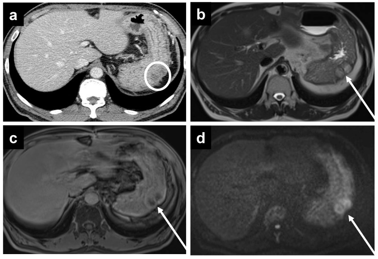
Figure 5.
A 67-year-old male with metastatic colonic adenocarcinoma and gastric serosa peritoneal deposits. The deposits are not as well seen on (a) CT, as denoted by the white circle, but are more apparent on MRI, including the (b) T2W, (c) delayed, and (d) DWI sequences secondary to the superior soft tissue contrast.

