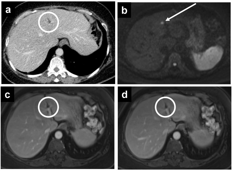
Figure 6.
A 61-year-old female with subtle recurrent primary peritoneal carcinoma, a tiny deposit is only well seen on the (b) DWI as a hyperintense focus at the falciform ligament (white arrow). A rim-enhancing nodule is barely seen on (a) CT, as well as the (c) portal venous and (d) delayed phases on MRI (white circles).

