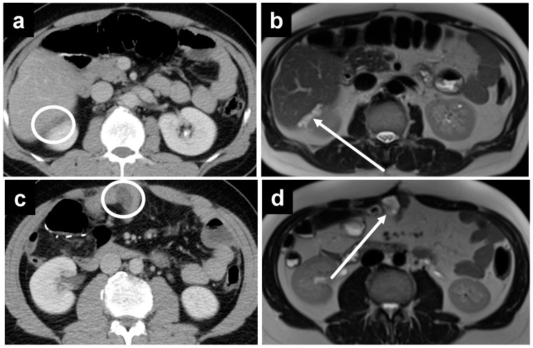
Figure 7.
A 44-year-old male with recurrent mucinous appendiceal tumor; the hypodense fluid is not well seen on the (a) CT at Morrison’s pouch but easily seen on the (b) T2W MRI, where the fluid also appears to be loculated, suggestive of a tumor deposit as opposed to bland ascites. Similarly, the pocket of fluid among small bowel loops is not as well appreciated on (c) CT but is well seen on (d) T2W MRI.

