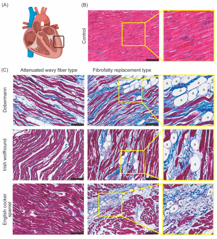
Figure 1.
Histologically distinct forms of canine DCM. Masson’s trichrome staining of 4 µm thick paraffin embedded slides [39]. (Red = cardiomyocytes, blue = fibrotic/connective tissue, a = adipocyte, black scale bar = 100 µm). (A) Model of the heart showing the location (depicted in the black box) of the collected tissue in the left ventricle. (B) Control tissue (3-year-old male Beagle with no cardiac symptoms) showing healthy cardiomyocytes. (C) Diseased tissue (4-year-old male Dobermann, 5-year-old Irish wolfhound, and 7-year-old English cocker spaniel) showing the attenuated wavy fiber type and fibrofatty type in three pathologically confirmed DCM cases.

