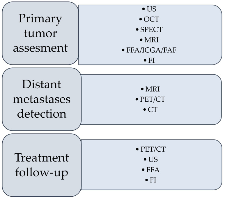
Figure 1.
Preferable imaging techniques in different stages of diagnosis and follow-up of uveal melanoma. US—ultrasonography; OCT—optical coherence tomography; SPECT—single-photon emission computed tomography; MRI—magnetic resonance imaging; FFA—fundus fluorescein angiography; ICGA—indocyanine green angiography; FAF—fundus autofluorescence; PET—positron emission tomography; CT—computed tomography.

