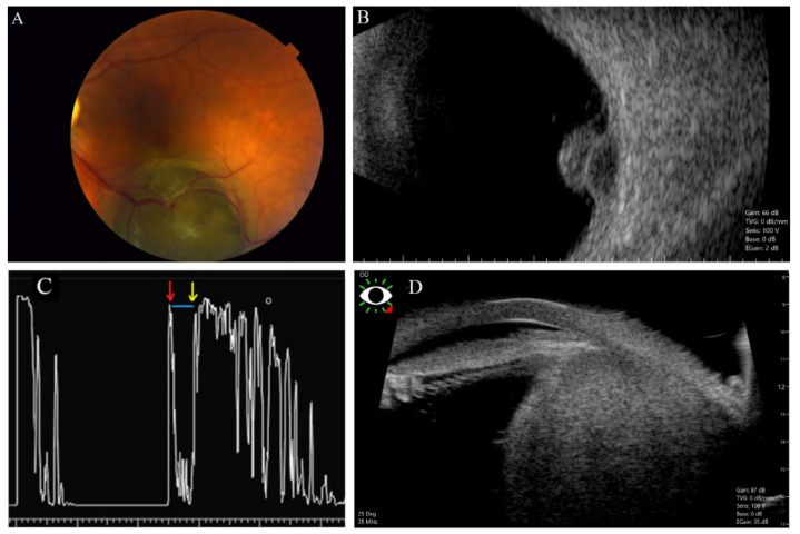
Figure 3.
Echographic features of choroidal melanoma. Melanotic choroidal melanoma is mushroom-shaped as detected at the level of inferior arcade of the left eye (A). That was shown to be mushroom-shaped with acoustic hollowness in B-scan (B). One-dimensional A-scan imaging (8 MHz) for an eye with choroidal melanoma (C). The vertical deflections represent echoes from different surfaces in the eye. The red arrow indicates the surface of the retina, the yellow arrow indicates the surface of the sclera, and the interval between them (blue line) shows the low reflectivity of the tumor (choroidal melanoma). Ciliary body melanoma (D) detected in ultrasound biomicroscopy (UBM).

