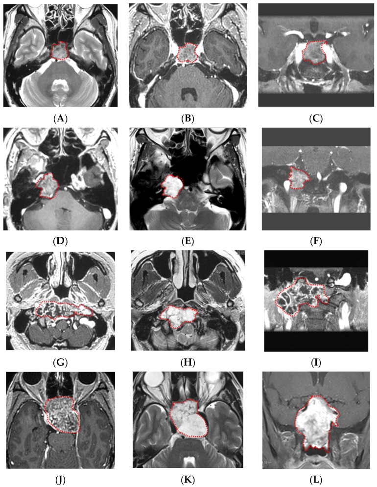
Figure 4.
Representative cases of chordomas and chondrosarcomas. (A–C) GdT1, T2, and fsGdT1 coronal images of a typical chordoma case. The machine learning model correctly diagnosed the tumor, and high diagnostic accuracy (80%) by neurosurgeons was noted. (D–F) GdT1, T2, and fsGdT1 coronal images of a typical chondrosarcoma case. The machine learning model correctly diagnosed the tumor, and the highest diagnostic accuracy by the neurosurgeons (85%) was noted. (G–I) GdT1, T2, and fsGdT1 coronal images of the chordoma case with the correct diagnosis by the machine learning model but with low diagnostic accuracy by the neurosurgeons (40%). (J–L) GdT1, T2, and fsGdT1 coronal images of the chondrosarcoma case with the incorrect diagnosis by the best machine learning model and the lowest diagnostic accuracy by the neurosurgeons (20%). Abbreviations: T2 weighted image—T2; post-gadolinium T1-weighted images—GdT1; fat suppression post-gadolinium T1-weighted images—fsGdT1. Red dotted lines indicates tumor.

