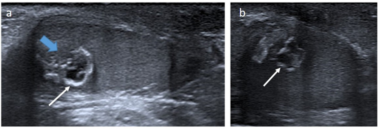
Figure 2.
Epidermoid cyst in a 20-month-old boy. Longitudinal (a) and transverse (b) gray-scale B mode US of the testis. Images show a well-demarcated intra-testicular lesion (blue arrow) with a small central anechoic part and a peripheral hyperechoic rim, giving an “onion-ring appearance” (white arrows). This lesion was incidentally discovered on scrotal US for a contralateral undescended testis. Serum levels of tumor markers were normal. The patient underwent conservative surgery. US, ultrasonography.

