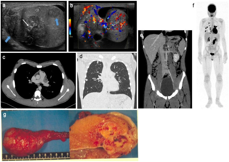
Figure 4.
Mixed non-seminomatous malignant germ cell tumor in a 14-year-old boy with yolk-sac and choriocarcinoma components. Longitudinal gray-scale B mode (a) and transverse color Doppler (b) US of the testis. Contrast-enhanced CT on axial view of the mediastinum (c), coronal view of the lung (d) and coronal view of the abdomen and pelvis (e). Coronal view of 18F-FDG PET (f). Images show several heterogeneous testicular lesions (blue arrow) containing micro calcifications (white arrow) and highly vascularized tissular portions with irregular distribution (b). CT scan demonstrates metastatic mediastinal and retroperitoneal lymph nodes and multiple lung metastases with high uptake on 18F-FDG PET. (g) Macroscopic view with gross pathology section of the lesion after radical surgery. This adolescent presented a rapid increase in testicular volume and increased AFP and BHCG levels. US, ultrasonography; AFP, alpha-fetoprotein; BHCG, beta-human chorionic gonadotrophin; 18F-FDG PET, 18F fluorodeoxyglucose positron emission tomography. Courtesy of Dr Brisse and Dr Cardoen, Institut Curie, Paris, France and Pr Berrebi, Robert Debré Hospital, Paris, France.

