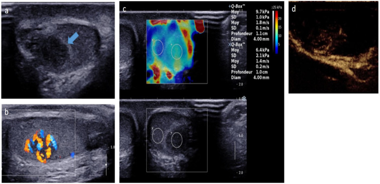
Figure 5.
Leydig-cell tumor in an 8-year-old boy. Transverse gray-scale B-mode (a) and longitudinal color Doppler (b) US of the testis. Shear wave elastography cartography (c) and contrast-enhanced US focused on the lesion (d). Images show a well-circumscribed hypoechoic mass with intrinsic central and peripheral hypervascularization, surrounded by a hyperechoic rim (blue arrow) with moderate increased stiffness on elastography. The patient was referred for early pubic hairiness and accelerated growth rate. Testosterone level was increased, and gonadotrophin levels (follicle-stimulating hormone, luteinizing hormone) were low. The patient underwent conservative surgery. US, ultrasonography. Courtesy of Pr Laurence Rocher, Antoine Béclère, Clamart, France.

