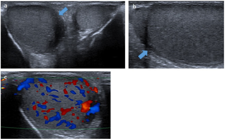
Figure 8.
Leukemic testicular infiltration of the right testis in a 10-year-old boy. Transverse (a) and longitudinal (b) gray-scale B-mode and transverse color Doppler (c) US of the testis. Images show an enlargement of the right testis demonstrating an ill-defined hypoechoic infiltration (blue arrow) with increased intralesional flow preserving the normal vascular architecture. This infiltration regressed on chemotherapy. US, ultrasonography.

