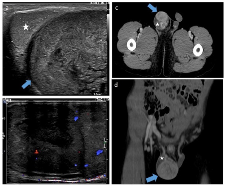
Figure 14.
Para-testicular rhabdomyosarcoma in a 6-year-old boy (case 2). Transverse gray-scale B-mode (a) and color Doppler (b) US of the scrotum. Staging contrast-enhanced CT scan focused on the pelvic on axial (c) and coronal (d) views. Images show a solid heterogeneous lesion (blue lesion) in an extra-testicular location (testis is represented by a star) with internal vessels on color Doppler images and showing high and heterogeneous enhancement after contrast administration. US, ultrasonography.

