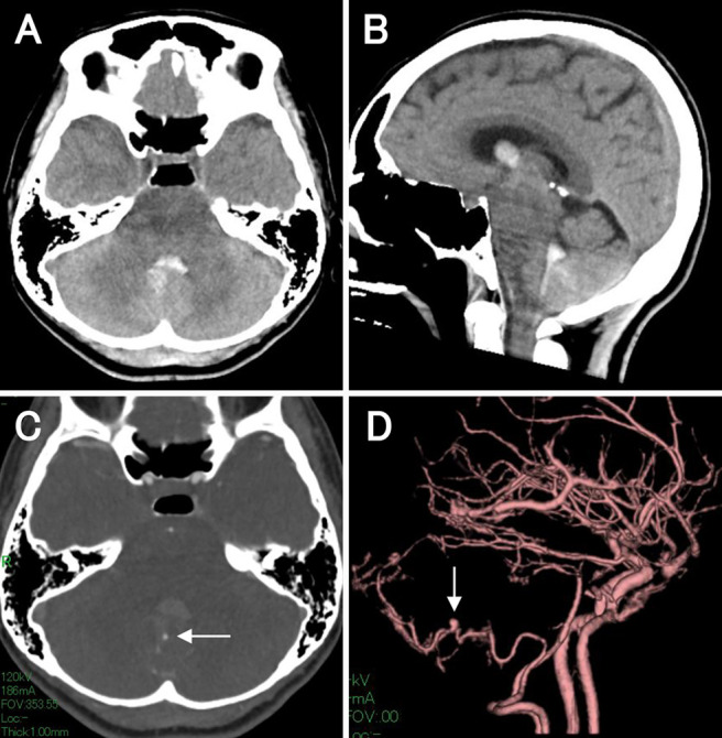
FIG. 1.

Initial noncontrasted CT and CTA at admission. Noncontrasted axial (A) and sagittal (B) CT showed a hematoma in the fourth ventricle. CTA axial image (C) and three-dimensional CTA (D) detected a small aneurysm (white arrows) that was located at the main trunk of the right PICA near the fourth intraventricular hemorrhage and was associated with the bleeding.
