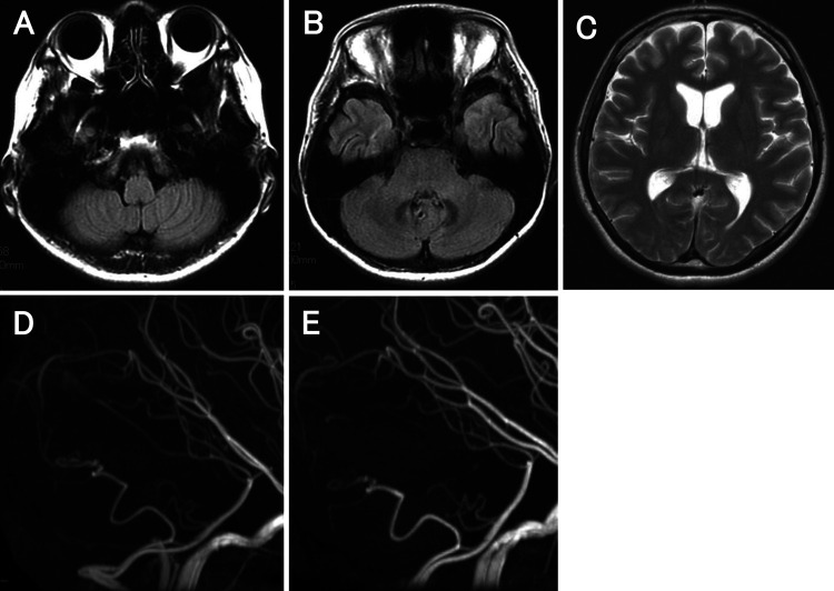
FIG. 5.
Follow-up MRI/MR angiography (MRA) after the treatment. A and B: Axial images of fluid-attenuated inversion recovery at 3 weeks after treatment showed no infarction in the bilateral cerebellar hemisphere. C: Axial T2-weighted image at 6 weeks after treatment showed a normal-sized ventricle without hydrocephalus. D: Lateral view MRA at 6 weeks after treatment showed no regrowth or recurrence of the coiled aneurysm. E: Lateral view MRA at 6 months after treatment revealed no regrowth or recurrence of the coiled aneurysm.

