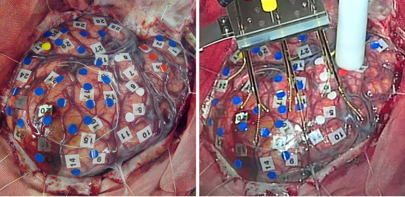
FIG. 4.
Intraoperative photograph of patient 2. Left: Functional mapping by electrical stimulation. The red, yellow, white, and blue markings indicate the motor and sensory language areas, motor cortex, and areas with no response to the stimulation, respectively. The area in the circle indicates the tumor location. Right: The cooling probe was attached to the brain surface. Spring electrodes were prepared for observation after the discharge.

