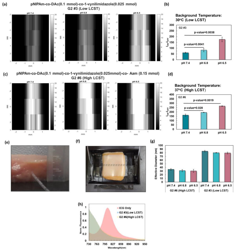
Figure 4.
(a) Imaging of the low LCST (30°C) polymeric nanoparticle of pNIPAm-co-DAc (0.1 mmol)-co-1-vynilimidazole (0.025 mmol), G2 #3, inside a silicone tube at the depth of ~1 cm in chicken tissue. (b) USF signal significantly increased from pH 7.4 (physiological pH), to 6.8 and 6.5 (higher and lower limits of tumor pH). (c) Imaging of pNIPAm-co-DAc (0.1 mmol)-co-1-vynilimidazole (0.025 mmol)-co-Aam (0.15 mmol) with high LCST of 38°C. (d) There was a significant difference in USF signal between pH 7.4 (physiological), compared to 6.8, and 6.5 (tumor pH). (e) and (f) Silicone tube inserted at the depth of ~1 cm in chicken tissue for USF imaging. (g) Effective diameter of samples G #3(Low LCST) and G #6 (High LCST) at 25°C.(h) Emission spectra of ICG and samples G #3 and G #6 at the excitation of 671 nm with an emission filter of 715 long-pass. (A color version of this figure is available in the online journal.)

