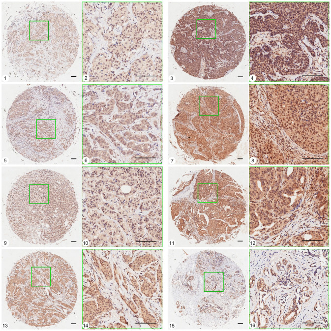
Figure 1.
Representative pictures of Pink1 expression in different subtypes of breast cancer tissues and breast adenosis using TMA-ISH method. 1, 2 and 3, 4: expression of Pink1 in basal-like subtype tissue, grade I (weak staining) and grade III (strong staining). 5, 6 and 7, 8: expression of Pink1 in HER2 over-expression subtype tissue, grade I (weak staining) and grade III (strong staining). 9, 10 and 11, 12: expression of Pink1 in Luminal B subtype tissue, grade I (weak staining) and grade III (strong staining). 13, 14 and 15, 16: expression of Pink1 in Luminal A subtype tissue, grade I–II and breast adenosis. (A color version of this figure is available in the online journal.)
Ruler scale = 100.8 μm.

