Figure 4.
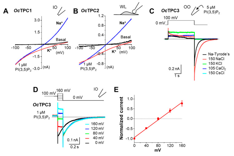
All three TPC subtypes from rabbit are Na+ selective when activated by PI(3,5)P2. HEK293 cells were transiently transfected with cDNA coding for OcTPC1-EGFP, OcTPC2-EGFP, or OcTPC3-EGFP. (A,B) Representative I–V curves derived from voltage ramps for OcTPC1 in an inside-out (IO) patch (A) and OcTPC2 in a whole-endolysosome (WL) patch (B) sequentially exposed to 1 µM PI(3,5)P2 in the normal bath solution containing mainly K+ (red traces) and then in a Na+-based bath solution (blue traces). (C) Representative current traces of OcTPC3 detected in an outside-out (OO) patch containing 5 µM PI(3,5)P2 in the pipette solution and sequentially exposed to normal Tyrode’s solution followed by equiosmotic solutions that contained 150 mM NaCl, 150 mM KCl, 105 mM CaCl2, and 150 mM CsCl. Note the lake of tail currents in KCl, CaCl2, and CsCl. (D) Representative current traces of OcTPC3 detected in an IO patch exposed to 1 µM PI(3,5)P2 in normal Tyrode’s solution. After stepping to +100 mV for 4 s, the voltage was switched to 0, 40, 80, 120, and 160 mV for 100 ms before returning to 0 mV. For clarity, only the last 0.1 s of the +100-mV step is shown together with the tail currents at post-step potentials of 0 to 160 mV followed by that at 0 mV. For (C,D), dashed lines indicate zero current. (E) I–V relationship of currents at the post-step potentials normalized to that of the absolute value at 0 mV for patches recorded as in (D). Data points are means ± SEM of n = 5 patches fit by linear regression.

