Figure 4.
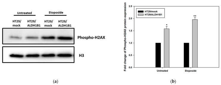
Evaluation of H2AX phosphorylation (serine 139) (phospho-H2AX) in HT29/ALDH1B1 and HT29/mock cells. (a) Forty (40) μg of HT29/ALDH1B1 and HT29/mock cell extracts were subjected to western blot analysis against H2AX phosphorylation. Histone H3 was used as a control for equal loading. (b) The protein expression levels of the phosphorylated (ser139) H2AX were >1.5-fold and >1.8-fold higher under normal and etoposide conditions, respectively, compared to HT29/mock cells. * p < 0.05, ** p < 0.01.

