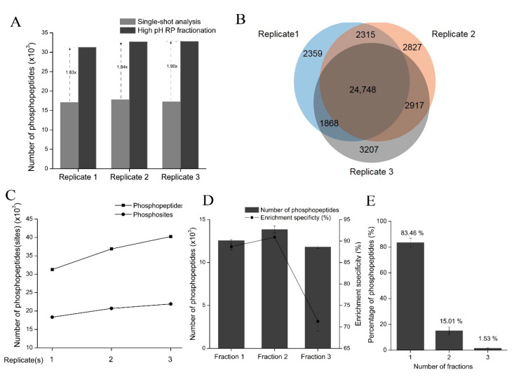
Figure 7.
The TEA-based HpH-RP fractionation of phosphopeptides. (A) Compared with single-shot LC-MS/MS analysis, HpH-RP fractionation increases the number of phosphopeptides identified from the lactic acid method by nearly two-fold. Three replicates are performed for both single-shot analysis and HpH-RP fractionation. (B) The overlap of the phosphopeptides identified in the three replicates of HPH-RP fractionation. (C) The cumulative number of phosphopeptides and phosphosites identified in 293T cells. (D) The number of phosphopeptides identified and phosphopeptide enrichment specificity in the three fractions of the TEA-based HpH-RP fractionation. Bars show the mean ± SD of three replicates; error bars indicate standard deviation. (E) The separation efficiency of the TEA-based HpH-RP fractionation is shown as the percentage of phosphopeptides found in one or more fractions.

