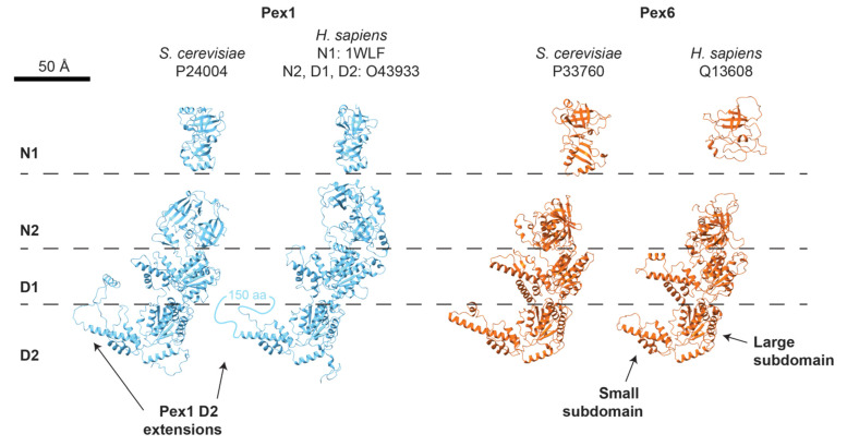
Figure 4.
Structures of Pex1 (blue) and Pex6 (orange) monomers from AlphaFold2 or, for HsPEX1 N1, X-ray crystallography (PDB 1WLF, [148]). N1 domains are separated for clarity; Pex1 N1 domains are flexibly attached to the motor, while Pex6 N1 domains are rigidly attached to the Pex6 D1 ring (see Figure 2).

