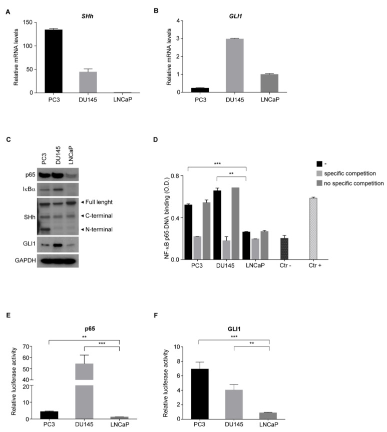
Figure 3.
NF-κB promotes the SHh pathway in AI PCa cell lines. (A,B) Real-time PCR showing basal level of SHh (A) and GLI1 (B) in AI (PC3, DU145) and AD (LNCaP) cell lines. (C) Western blots showing the basal level of p65, IκBα, SHh and GLI1 proteins in AI and AD cell lines. GAPDH is shown as the loading control. (D) TransAM assay in AI (PC3, DU145) and AD (LNCaP) cell lines. (E,F) Dual luciferase reporter assay for p65 (E) and GLI1 (F) in AI and AD cell lines used in (A). Luciferase activity was normalized to Renilla activity. All experiments were performed in triplicate. Values are shown as mean ± S.D. Unpaired Student t-test or Mann–Whitney U-test were used for statistical analysis. ** p < 0.01; *** p < 0.001.

