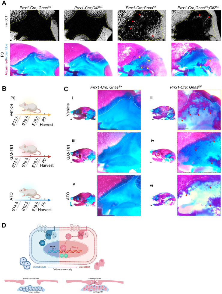
Figure 5.
Reduction of Gli transcription activity largely rescued cartilage ossification. (A) Upper: micro–computed tomography 3-dimensional image of Prrx1-Cre; Gnasfl/fl; Gli2fl/+ mouse and the control littermate calvarias. Dotted lines outline enlarged view of mastoid fontanel. The arrows (red) indicate reduced heterotopic ossification (HO). Scale bars: 1 mm. Lower: skeletal preparation of mouse calvarias and their control littermates. The arrows (yellow) indicate reduced HO in rescue group. Scale bar: 1 mm. (B) Schematics of experiment design. (C) Skeletal preparation of mouse calvarias receiving GANT61, ATO, or DMSO (vehicle). The arrows (red) indicate reduced HO. Scale bar: 1 mm. (D) Schematics of this work: a Gαs-regulated conversion of cranial chondrocyte identity through Hedgehog signaling strengthens HO during calvaria development.

