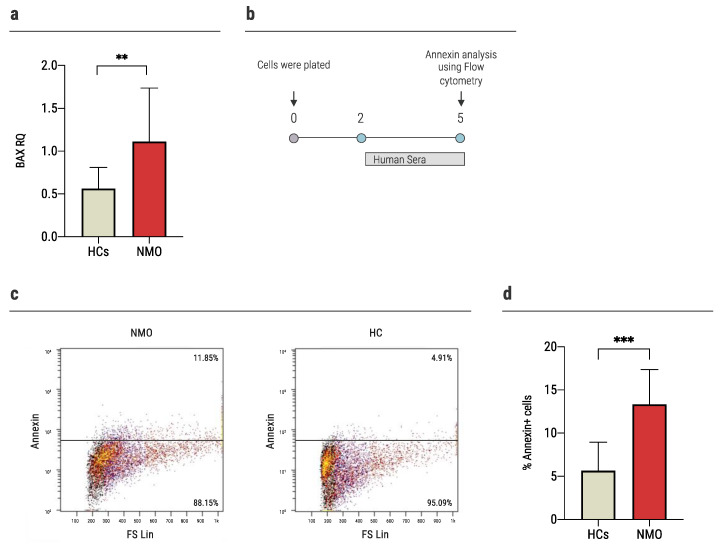
Figure 4.
Exposure of NMO sera increased apoptosis in astrocytes. (a) The expression of BAX increased significantly in the astrocytes cultured with sera of NMO patients (1.11 ± 0.62, n = 17) vs. 0.56 ± 0.24 (n = 13) of the HCs, as was determined using rt-QPCR, (b) time-course experiments of annexin levels. Primary astrocytes were cultured with human sera (20% of media) for 72 h, followed by evaluation of apoptosis expression using flow cytometry, (c) representative flow cytometry analysis of apoptotic astrocytes upon culture with sera obtained from seropositive NMO patients or HCs, (d) higher levels of apoptosis among astrocytes cultured with sera of NMO patients (13.34 ± 4.03%, n = 10), compared to HCs (5.7 ± 3.3%, n = 10), as evaluated using annexin staining. Each assay was repeated independently at least three times. Significance was determined by unpaired two-tailed student’s t-test (p ** ≤ 0.01, *** ≤ 0.001). Error bars in all graphs represent standard deviation.

