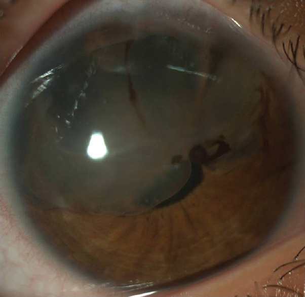
Figure 5.

Slit-lamp photograph (25X magnification) of the right eye showing clear fluid filled, irregular, isolated, loculated, translucent iris cyst present from 9 to 2 o’clock position covering the pupillary axis attached to the corneal endothelium with no variable pigmentation/abnormal vasculature with dense iris pigments inside it
