Figure 7.

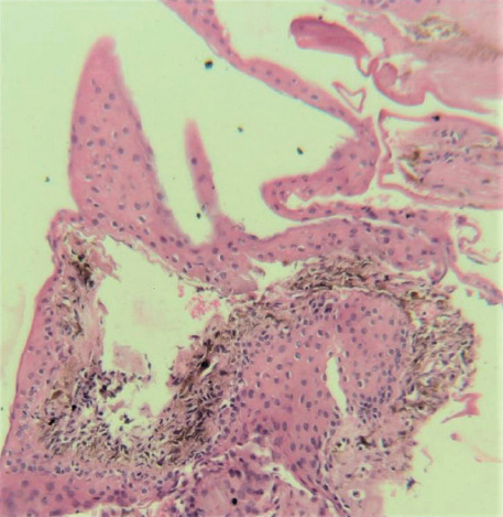
Histopathology image (10X magnification) showing iris cyst lined with normal squamous epithelium with no dysplasia or atypia with interspersed melanocytes. Goblet cells or malignant cells were not seen

Histopathology image (10X magnification) showing iris cyst lined with normal squamous epithelium with no dysplasia or atypia with interspersed melanocytes. Goblet cells or malignant cells were not seen