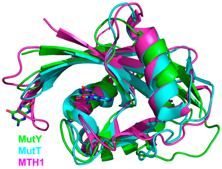
Figure 4.
Overlay of NUDIX domains from G. stearothermophilus MutY (green; PDB ID 1RRQ [138]), E. coli MutT (cyan, PDB ID 3A6T [144]), and human MTH1 (magenta, PDB ID 3ZR0 [145]). OxoG bases at their respective binding sites are shown as stick models with carbon atoms colored the same as the respective protein.

