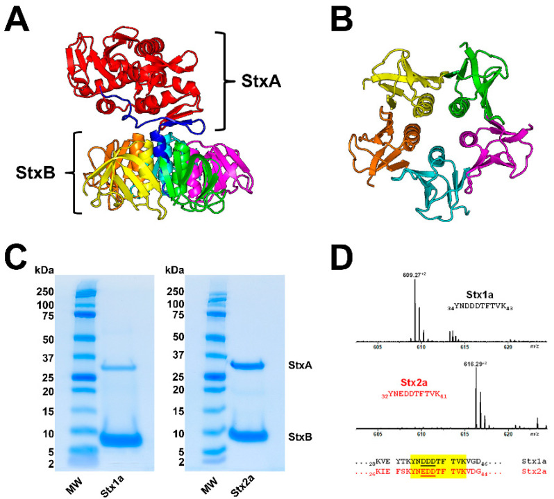
Figure 1.
Ribbon diagrams of the Stx2a crystal structure (A) and the homopentameric StxB viewed from below (B); SDS-PAGE-separated affinity-purified Stx1a and Stx2a (C) and signals of Stx1a and Stx2a subtype-specific diagnostic peptide ions detected by mass spectrometry (D). The structure of Stx2a (1R4Q) was drawn with PyMOL Molecular Graphics System, Version 2.0 (Schrödinger, Synaptic Science, LLC, Bethesda, MD, USA) based on the amino acid sequence published by Fraser and co-workers [171] deposited in the Research Collaboratory for Structural Bioinformatics (RCBS) data bank (https://www.rcsb.org/structure/1R4Q, 5 June 2022). The StxA subunits (StxA) and StxB subunits (StxB) were separated under reducing conditions and stained with Coomassie Blue (C). Diagnostic peptide ions derived from tryptic digestions of the B subunits of Stx1a and Stx2a, the peptide positions and their amino acid sequences within the B subunits are highlighted in yellow (taken from Steil and co-workers [170].

