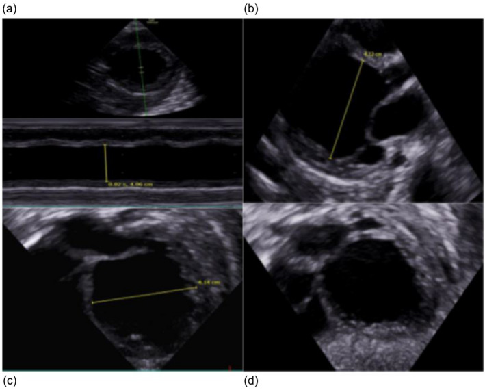
FIGURE 2.

Echocardiography shows dilated cardiomyopathy in our patient at 5 months. (a) Left ventricle (LV) m mode showing severe reduced contractility and LV dilatation. (b) PLAX 1: End diastole. (c) PLAX 2: End diastole LV diameter. (d) Subcostal 4ch: End diastole showing very dilated LV.
