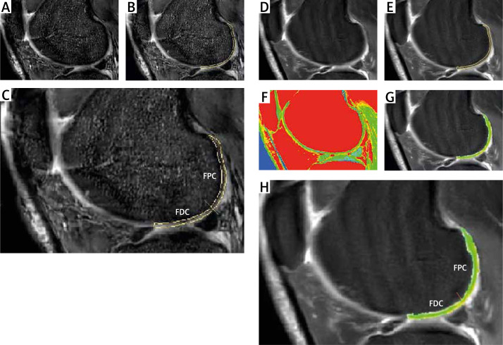
Figure 1.
T1 rho/T2 mapping and ROIs. A – Sagittal T1rho sequence was performed. B – The regions of interest (ROI) were drawn. The posterior edge of the anterior meniscal horn was used to separate the edge of ROI. C – ROI of condyles was divided into two parts: the femoral distal condyle and the femoral posterior condyle. The posterior edge of the posterior meniscal horn was used to segment the femoral distal condyle. D – T2 Fast spin-echo proton density-weighted fat suppression imaging (FSE PDFs) was performed. E – ROI of T2 was drawn. F – A sagittal T2 mapping sequence was performed. G – T2 images were merged with the ROI of T2 mapping images. H – The femoral distal condyle and the femoral posterior condyle were outlined

