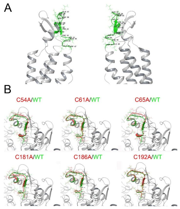
Figure 6.
MD simulations suggest changes in the 3D disposition of the TM1-EL1 parahelix segment. Human Cx46 50-ns MD models. (A) Representation of a Cx46 hemichannel showing the location of the parahelix (green segment). (B) Zoom of the parahelix segment (the segment between amino acids 47 and 61 was analyzed). The configuration of this segment in the wild-type Cx46 (green) is superimposed on the parahelix model for each Cys mutant (red).

