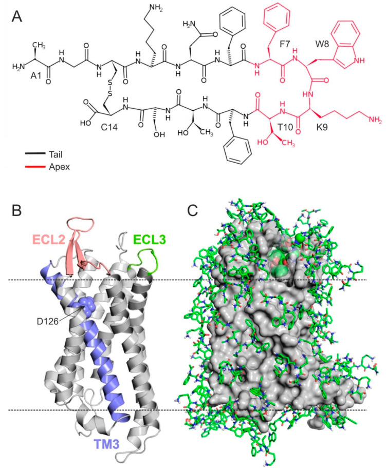
Figure 1.
(A) Lewis structure of SST highlighting the apical FWKT region with red. (B) Homology model of SSTR4 in cartoon representation. D126 (spheres) on TM3 (teal), ECL2 (salmon), and ECL3 (green) are proved to be important in ligand binding and receptor activation. (C) SSTR4 (grey, surface) covered with monolayer of tetrapeptide fragment (Ace–FWKT–NHMe) copies (green, all atom, sticks) at the end of the 7th docking cycle. The best energy fragment is highlighted with spheres (green, all atom).

