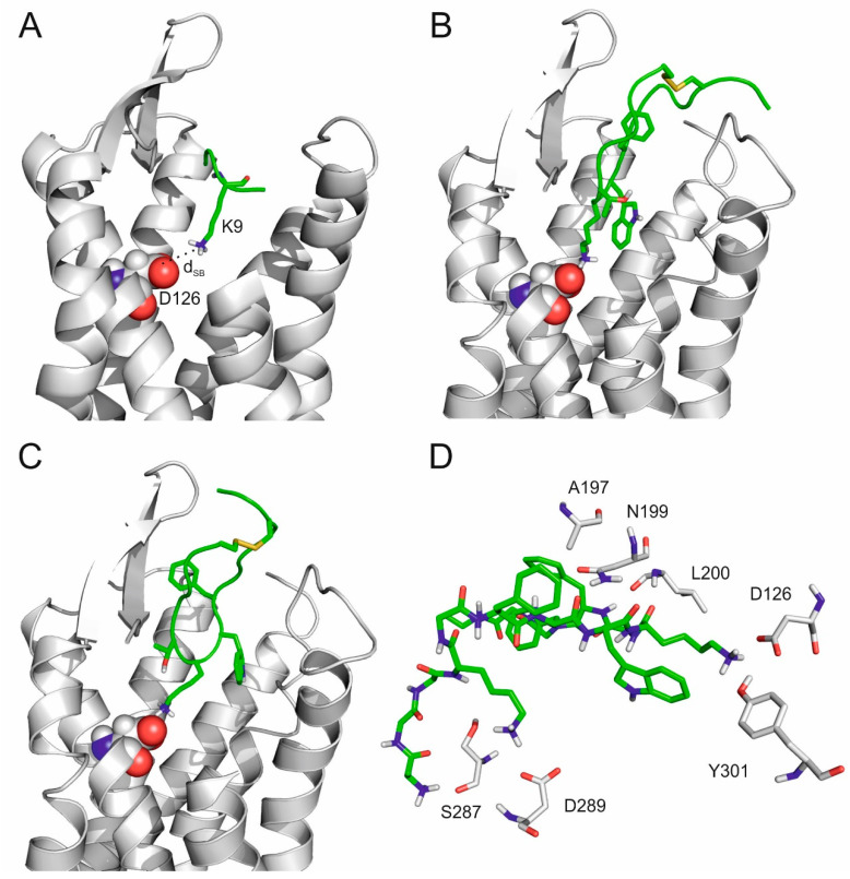
Figure 4.
(A) Complex of SSTR4 (grey, cartoon, D126 highlighted by spheres) and the tetrapeptide fragment (green, K9 highlighted by sticks) with the smallest dSB in the 100 ns-long MD simulation. (B,C) Internal binding mode of SST with 3.1 Å (B) and 3.0 Å dSB determined in separate 350 ns-long MD simulations. (D) The close-up view of SST (green, sticks) in the internal binding mode surrounded with target residues (grey, sticks) within a 3.5 Å distance from the ligand.

