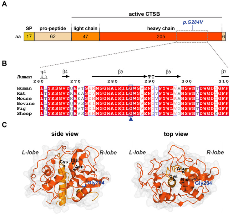
Figure 3.
Glycine 284 is conserved in CTSB across species. (A) Primary structure of CTSB indicating length of fragments in amino acids (aa) and position of the p.Gly284V mutation. SP—signal peptide. (B) Structure-informed multiple sequence alignment of six CTSB homologs. The secondary structure for human CTSB (PDB: 6AY2) is shown above. Boxed residues are conserved: white background with red text indicates functionally equivalent residues; red background with white text indicates sequence conservation. The blue box and arrowhead highlight Gly284, which is conserved across all species analyzed. (C) Crystal structure of mature human CTSB (PDB: 6AY2) shown in surface/ribbon representation, indicating the left (L) and right (R) lobes which constitute the mature enzyme. Heavy and light chains are colored red-orange and orange, respectively. Gly284 (blue) is shown in cylindrical representation; C-alphas of the catalytic triad: Cys (29) His (199) and Asn (219) are depicted as black spheres.

