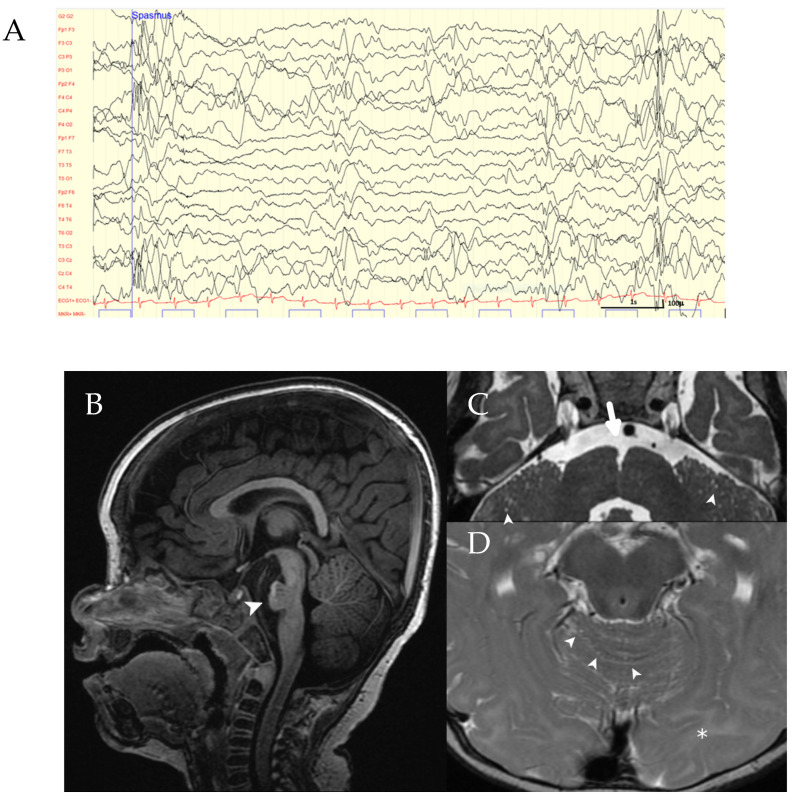
Figure 1.
Clinical findings in the index patient. (A) EEG of the patient showing generalized epileptiform discharges. (B–D) Brain MRI: Sagittal T1-weighted image showing pontine hypoplasia (arrowhead in (B)). Axial T2-weighted images (C,D) demonstrating an abnormally marked anterior median fissure at the pontomedullary junction (large arrow in (C)), multiple microcysts in the cerebellar hemispheres and cerebellar vermis (arrowheads in (C,D)), and hyperintense signal of the occipital white matter (asterisk in (D)).

