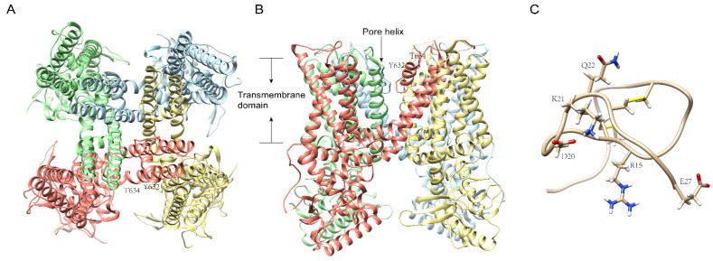
Figure 4.
The cryo-electron microscopy structure of TRPV1 and RhTx. Ribbon diagram of TRPV1 atomic model (PDB id: 7L2L) with each of the four identical subunits color-coded, showing views from the bottom (A) and side (B). The key residues for the interaction with RhTx are labeled. The membrane-spanning helices and different subunits of the TRPV1 channel are indicated. (C): RhTx from Scolopendra subspinipes. PDB id: 2MVA.

