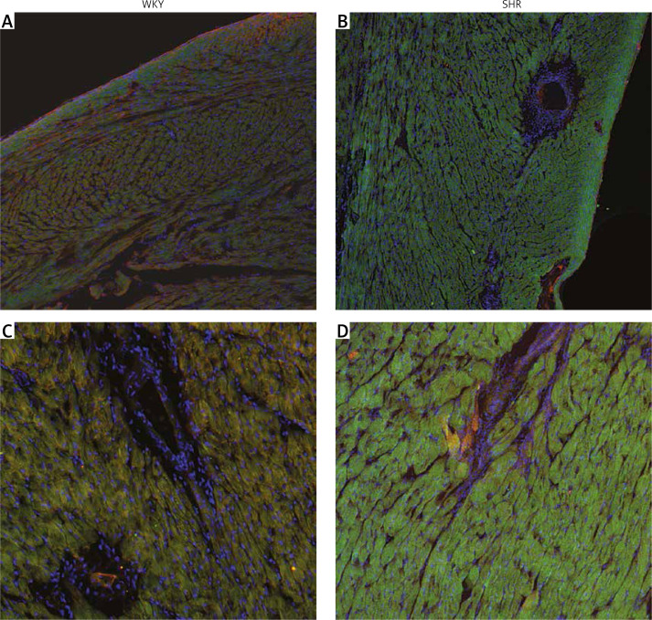
Figure 3.
Immunostaining of the cardiac muscle of the left ventricle in the normotensive Wistar-Kyoto (WKY; A, C) and in spontaneously hypertensive (SHR) rats (B, D). Figure shows hypertrophy of the left ventricle cardiomyocytes and high intensity of oxytocin receptor (OTR; green staining) in the myocytes of the left ventricle in the SHR rats. Staining of the cell nuclei with Hoechst 33258 (blue), oxytocin receptor OTR (green), and α-skeletal muscle actin – aSMA (red). The bar – 100 μm

