Figure 1.
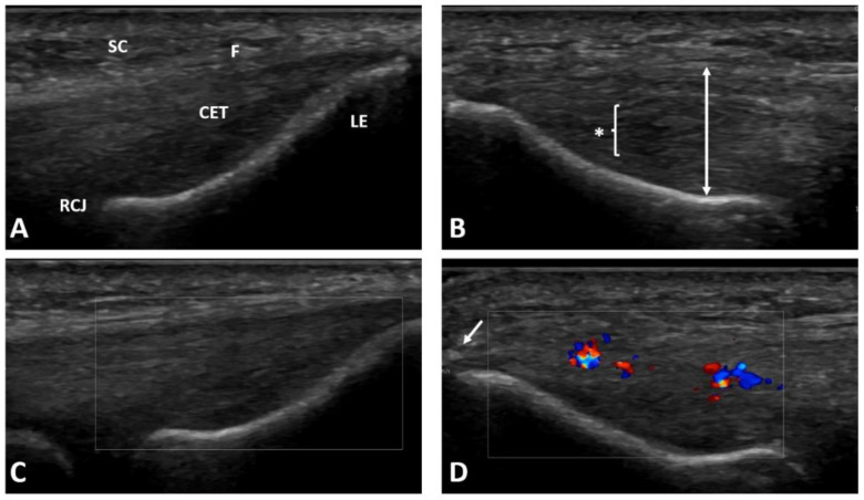
Ultrasound examination of the patient’s both elbows for lateral elbow tendinopathy confirmation: (A) healthy lateral epicondyle region of the contralateral elbow; (B) pathological common extensor tendon thickening and region with diffuse hypoechogenicity typical for lateral elbow tendinopathy; (C) healthy common extensor tendon without activity in color Doppler; (D) neoangiogenesis shown as increased activity in color Doppler in the tendinopathic region. SC—subcutaneous tissue; F—fascia; CET—common extensor tendon; LE—lateral epicondyle; RCJ—radiocapitellar joint; * and bracket—diffuse hypoechogenic region; two-head arrow—tendon thickness measurement; arrow—enthesophyte.

