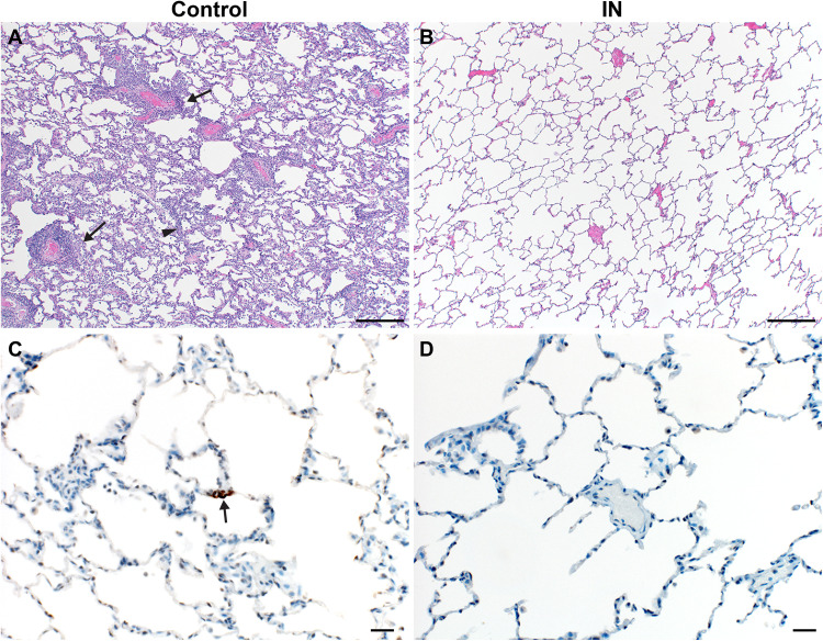
Fig. 7. Lung pathology is reduced in ChAdOx1 nCoV-19 IN-vaccinated rhesus macaques after SARS-CoV-2 challenge.
(A and B) Lung tissue sections isolated from IN-vaccinated (A) and control (B) rhesus macaques were stained with H&E. Scale bars, 200 μm. (A) Interstitial pneumonia (arrowhead) and lymphocytic perivascular cuffing (arrows) were observed in control samples. (B) No pathology was observed in IN-vaccinated lung samples. (C and D) Immunohistochemistry for SARS-CoV-2 antigen (brown) reveals rare type I pneumocyte immunoreactivity (arrow) in control samples (C) but no immunoreactivity in IN-vaccinated samples (D). Scale bars, 20 μm.

