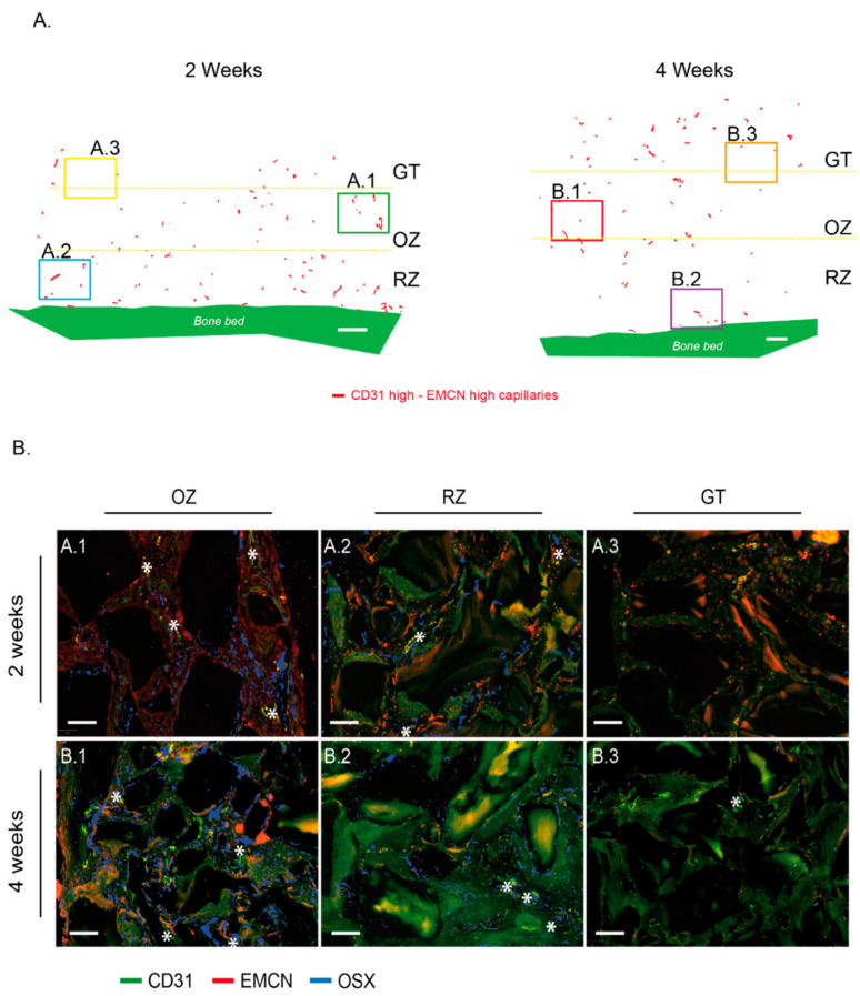
Figure 6.
“CD31 High/EMCN High” capillaries distribution within the cylinder contents at 2 and 4 weeks and correlation with the presence of Osterix-expressing osteogenic precursors. (A) CD31 High/EMCN High capillaries from a representative image of a cylinder’s entire content at 2 weeks (left) and 4 weeks (right) are shown on a projection where tissues were digitally removed. The same representative sample was conserved from Figure 2A and Figure 3A, and Figure 5D at 2 weeks, Figure 2B and Figure 3B, and Figure 5D at 4 weeks. Granulation tissue (GT), osteogenic zone (OZ), and remodeling zone (RZ) were delimited by yellow dotted lines. White bar: 500 µm. (B) Representative magnifications from (A)—cylinder content at 2 weeks: OZ (A.1), RZ (A.2), and GT (A.3) and (B)—cylinder content at 4 weeks: OZ (B.1), RZ (B.2), and GT (B.3). Samples were analyzed by RNA hybridization for CD31 (green), EMCN (red), and OSX (blue). The “CD31 High/EMCN High” capillaries are tagged by a white star. Note that OSX-expressing osteogenic precursors were mainly observed in the osteogenic zone, in the vicinity of “CD31 High/EMCN High” rich regions, both at 2 (A.1) or 4 weeks (B.1). Despite the presence of “CD31 High/EMCN High” capillaries in the RZ, such concentrations of osteogenic precursors were not observed at 2 (A.2) or 4 weeks (B.2). A very weak expression of OSX was observed in the GT zone at 4 weeks (B.3), suggesting that osteogenic precursors migrate towards the region where “CD31 High/EMCN High” capillaries were observed. White bars: 100 µm.

