

Currarino Silverman syndrome is a rare form of pectus carinatum that was first described by Guido Currarino and Frederic Silverman in 1958. Pectus carinatum, along with pectus excavatum are the two most common congenital anterior chest wall deformities. Compared to pectus excavatum, pectus carinatum is present in only 5%–15% of patients with anterior chest wall abnormalities. There are two principal subtypes of pectus carinatum: Chondrogladiolar (type 1) and chondromanubrial (type 2). While the etiology is not fully understood, it is thought that the sternal deformity is due to an excessive growth of costal cartilage [1]. Type 2, also known as Currarino Silverman (CS) syndrome, is a developmental anomaly characterized by prominent sternal angulation and a decreased length due to complete, non-segmentation or premature synostosis of the ossification centers in the sternum [2]. CS syndrome is separated from other types of pectus carinatum by a unique appearance: a short, “Z” shaped sternum with prominent outward protrusion. A bilateral deformity of the second to fifth costal cartilages is also present and the xiphoid process is absent (Figure 1).
Figure 1.
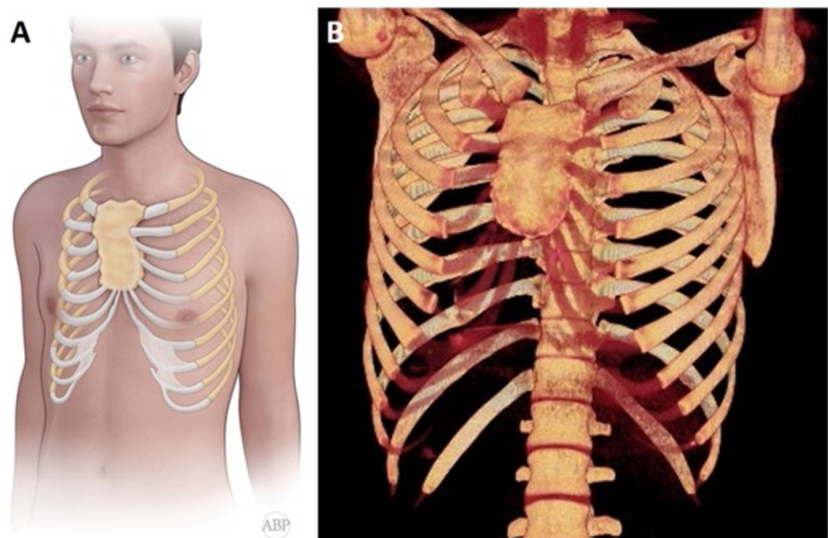
Anatomical representation of Currarino-Silverman syndrome: (A) short, broad, fused sternum with prominent outward protrusion; the xiphoid process is absent; deformed, fused and elongated costal cartilages formed abnormal sharp angle; (B) Chest CT scan with 3-D reconstruction.
In addition to unique physical features, CS syndrome is frequently associated with congenital heart defects. In 1956, Monnet et al. [3] documented the first case of congenital heart disease associated with a sternum that was entirely fused and angulated. During cardiogenesis, abnormal differentiation results in defects of the sternum, endocardial cushion, and aortic arch derivatives [4]. These embryologic changes may manifest as patent ductus arteriosus, atrial and/or ventricular septal defects, tetralogy of Fallot, transposition of the great arteries, and coarctation of the aorta. CS syndrome is also often associated with spinal deformities as well as both Turner’s syndrome and Noonan’s syndrome [5,6]. In our case series, echocardiography revealed various cardiac pathology in three of four patients: mitral and tricuspid regurgitation, mitral valve prolapse, and ventricular septal defect [7].
CS syndrome is commonly confused with pectus excavatum as both deformities appear similarly, but the surgical treatment approaches are very different. Proper classification is the foundation of appropriate surgical treatment. Acastello [8] differentiated various chest wall deformities based on the origin of the defect: type 1 (cartilaginous), type 2 (costal), type 3 (chondrocostal), type 4 (sternal) and type 5 (clavicle-scapular). CS syndrome was classified as type 1, or cartilaginous. Torre et al. [9] furthered the classification of type 2 pectus carinatum, stating that this sternal type was of normal length without depression of the lower third. On the other hand, a short, “Z”-shaped sternum pointed to CS syndrome. Patients with mild to moderate forms of CS syndrome can present with depression of the lower third of the sternum, attributing to confusion with pectus excavatum. In pectus excavatum, the sternal depression starts at the angle of Louis and deepens towards the xiphoid process along with elongated cartilages [10]. Thus, confusion about classification is still an actual issue. Terms “Currarino Silverman syndrome,” “pouter pigeon chest,” “chondromanubrial deformity,” “type 2 pectus carinatum” and “pectus arcuatum” are used, at times, interchangeably.
In 1952, Ravitch et al. [11] described his surgical approach to CS syndrome, which included two transverse osteotomies at the area of greatest sternal protrusion following elevation of the pectoralis major muscle and removal of deformed costal cartilage segments. Later, in 1960, he developed three main steps in the surgical treatment of CS syndrome: resection of the subperichondrial cartilage, preservation of the posterior sternal plate and periosteum with transverse sternotomy, and fixation of the chest wall shape, allowing for osteosynthesis [12].
In 1963, Robichek et al. [13] described his methods, which depended on the CS syndrome type: type A or type B. Type A (moderate depression of the sternum with manubrial protrusion and two adjacent cartilages) correction included subperichondrial resection of the protruding cartilage, transverse osteotomy, and anterior resection of the prominent manubrium. Type B (significant depression) correction included an upper level transverse wedge sternotomy at the depression with Marlex mesh placement.
Lam et al. [5] described a technique which included a 2 cm resection of the sternum at the most prominent point with two additional transverse osteotomies: one above and one below the resection. Brichon et al. [14] described a similar technique which included three osteotomies with resection of the curved anterior angle of the sternum. For chest wall stabilization, a Strasbourg Thoracic Osteosynthesis System (STRATOS) was placed in front of the sternum instead of bar placement. In another study, Kara et al. [15], used two STRATOS devices, which were introduced through the retrosternal space.
Fonkalsrud at al. [16] published a technique in which costal cartilages were reattached to the sternum and ribs, while preserving the perichondral sheath. An Adkins strut was placed posterior to the lower aspect of the sternum. This allowed for correction of sternal depression as well as preventing flail chest and paradoxical respiration postoperatively.
For stabilization, Kuzmichev et al. [17] described the use of parallel vertical titanium places placed on the inferior sternum, which provided for sternal osteosynthesis. Furthermore, Leng et al. [18] also described the use of vertical titanium plates. However, they utilized the use of technology based templates for design and osteotomy angle creation.
In a recent case series, we described modified Ravich technique with one and two level osteotomies using parallel stainless steel surgical monofilament wires and parallel low profile titanium plates for osteosynthesis of the sternum [7].
Due to the rare nature of CS syndrome and small number of publications surrounding surgical correction, no uniformly accepted surgical treatment has been described by current literature. However, Ravitch type procedure with multi-level wedge osteotomy remains the best surgical option and proper classification of CS syndrome is a mainstay of successful surgical repair.
Footnotes
Conflicts of Interest
No conflict of interest.
References
- 1.Fokin AA, Steuerwald NM, Ahrens WA, Allen KE (2009) Anatomical, histologic, and genetic characteristics of congenital chest wall deformities. Seminars in Thoracic and Cardiovascular Surgery 21(1):44–57. [DOI] [PubMed] [Google Scholar]
- 2.Currarino G, Silverman FN (1958) Premature obliteration of the sternal sutures and pigeon-breast deformity. Radiology 70(4):532–40. [DOI] [PubMed] [Google Scholar]
- 3.Monnet P, Gravier J, Gauthier J, Verney R (1956) Combination of partial vascular transposition (aortic dextroversion) with abnormal venous return and a thoracic malformation caused by premature ossification of the sternum. Pediatrie 11(1):95. [PubMed] [Google Scholar]
- 4.Gabrielsen TO, Ladyman GH (1963) Early closure of the sternal sutures and congenital heart disease. The American j roentgenol, radium ther nuc med 89:975–83. [PubMed] [Google Scholar]
- 5.Lam CR, Taber RE (1971) Surgical Treatment of pectus carinatum. Arch Surg 103(2):191–4. [DOI] [PubMed] [Google Scholar]
- 6.Chidambaram B, Mehta AV (1992) Currarino-Silverman Syndrome (Pectus Carinatum Type 2 Deformity) and Mitral Valve Disease. Chest 102(3):780–2. [DOI] [PubMed] [Google Scholar]
- 7.Gritsiuta AI, Bracken A, Beebe K, Pechetov AA (2021) Currarino-Silverman syndrome: diagnosis and treatment of rare chest wall deformity, a case series. J Thorac Dis 13(5): 2968–78. [DOI] [PMC free article] [PubMed] [Google Scholar]
- 8.Acastello E (2006) Patologias de la pared toracica en pediatria. Buenos Aires: Editorial El Ateneo. [Google Scholar]
- 9.Torre M, Rapuzzi G, Jasonni V, Varela P (2012) Chest Wall Deformities: An Overview on Classification and Surgical Options. InTech 117–36. [Google Scholar]
- 10.Fonkalsrud EW, Anselmo DM (2004) Less extensive techniques for repair of pectus carinatum: The undertreated chest deformity. J Amer College of Surg 198(6):898–905. [DOI] [PubMed] [Google Scholar]
- 11.Ravitch MM (1952) Unusual sternal deformity with cardiac symptoms operative correction. J of thor surg 23(2):138. [PubMed] [Google Scholar]
- 12.Ravitch MM (1960) Operative correction of pectus carinatum (Pigeon Breast). Annals of surgery 151(5):705–14. [DOI] [PMC free article] [PubMed] [Google Scholar]
- 13.Robicsek F, Daugherty HK, Mullen DC (1974) Technical considerations in the surgical management of pectus excavatum and carinatum. The Annals of Thoracic Surgery 18(6):549–64. [DOI] [PubMed] [Google Scholar]
- 14.Brichon P, Wihlm J (2010) Correction of a Severe pouter pigeon breast by triple sternal osteotomy with a novel titanium rib bridge fixation. Annals of Thoracic Surgery 90(6):e97–9. [DOI] [PubMed] [Google Scholar]
- 15.Kara M, Gundogdu AG, Kadioglu SZ, Cayirci EC, Taskin N (2016) The use of sternal wedge osteotomy in pectus surgery: When is it necessary? Asian Cardiovascular and Thoracic Annals 24(7):658–62. [DOI] [PubMed] [Google Scholar]
- 16.Fonkalsrud EW, Beanes S (2001) Surgical management of pectus carinatum: 30 Years’ Experience. World J Surg 25(7):898–903. [DOI] [PubMed] [Google Scholar]
- 17.Kuzmichev V, Ershova K, Adamyan R (2016) Surgical correction of pectus arcuatum. J visualized surg 2:55. [DOI] [PMC free article] [PubMed] [Google Scholar]
- 18.Leng S, Bici K, Facchini F (2019) Customized cutting template to assist sternotomy in pectus arcuatum. The Annals of Thorac Surg 107(4):1253–8. [DOI] [PubMed] [Google Scholar]