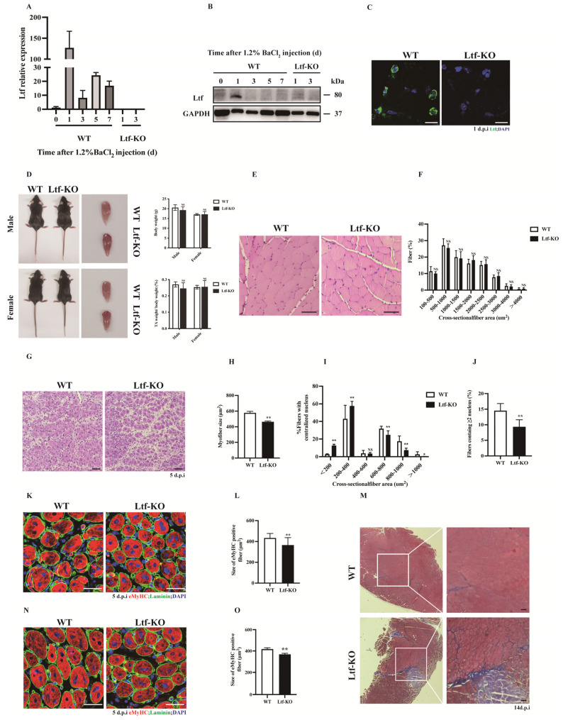
Figure 1.
Ltf deficiency impairs regenerative capability of skeletal muscle. (A) Real-time RT-PCR of Ltf expression levels in WT mice of TA muscle from days 0 to day 7 after BaCl2 injury and detected the TA tissue of Ltf-KO mice injured for 1 day and 3 days. (B) Immunoblots illustrate the protein levels of Ltf and an unrelated GAPDH in TA muscles from days 0 to day 7 after BaCl2 injury and detected the TA tissue of Ltf-KO mice injured for 1 day and 3 days. (C) Immunofluorescence results represent Ltf expression in WT and Ltf-KO mice of TA tissue on day 1 of injury. Scale bar: 10 μm. (D) Absolute body weight of adult WT and Ltf-KO mice, relative uninjured TA muscle wet weight of adult WT and Ltf-KO mice. (E) Pathological analysis plots represent hematoxylin and eosin (H&E) –stained TA cross-sections (CSA) of WT and Ltf-KO mice in the uninjured state. Scale bar: 60 μm. (F) Myofiber size (percentage) distributions of WT and Ltf-KO TA muscle were measured by using ImageJ software. (G) Representative photomicrographs of H&E-stained sections showing a delayed regeneration of injured TA muscle in Ltf-KO mice compared with that in WT at day 5 after BaCl2 injection. Scale bar: 60 μm. (H) Statistical analysis of mean area of TA muscle CSA in adult Ltf-KO and WT mice 5 days after injury. (I) Regenerated myofiber size (percentage) distributions of WT and Ltf-KO TA muscle 5 days after BaCl2 injury were measured by using ImageJ software. Only myofibers with centrally located nuclei were counted. (J) Quantification of the ratio of regenerating myofibers containing two or more centralized nuclei per field at day 5 post-injury. (K) Representative overlaid photomicrographs of TA muscle sections of WT and Ltf-KO mice 5 days post-injury after immunostaining for eMyHC (red) and laminin (green). Nuclei were labeled by DAPI. Scale bar: 30 μm. (L) Average CSA of eMyHC-positive fibers in TA muscle 5 days post-injury. (M) Representative photomicrographs of Masson-stained sections show increased collagen fibers in damaged TA muscles in Ltf-KO mice compared to WT at day 14 after BaCl2 injection. Scale bar: 60 μm. (N) At 60 days after the first injury, a second injection of BaCl2 solution was delivered to the muscle of WT and Ltf-KO mice, and the muscle was analyzed at day 5 after the second injury. Scale bar: 30 μm. (O) Average CSA of eMyHC-positive fibers in TA muscle at 5 days after the second injury. n = 3 in each group. * p< 0.05; ** p < 0.01; NS, not significant.

