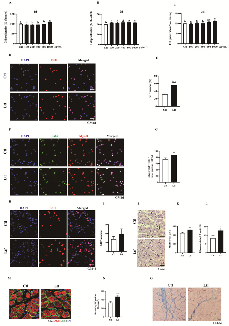
Figure 4.
R-Ltf promotes proliferation of SCs in vivo and in vitro. (A) Satellite cells (SCs) isolated from wild-type (WT) mice after 1 day of adherent growth, treated with various concentrations of R-Ltf for 1 day, and then added CCK-8 to determine the OD value. (B) SCs isolated from WT mice after 1 day of adherent growth, treated with various concentrations of R-Ltf for 2 days, and then added CCK-8 to determine the OD value. (C) SCs isolated from WT mice after 1 day of adherent growth, treated with various concentrations of R-Ltf for 3 days, and then added CCK-8 to determine the OD value. (D) SCs isolated from WT mice after 1 day of adherent growth, treated with 1000 μg/mL R-Ltf for other 3 days. Cells were labeled with EdU and DAPI. Scale bar: 20 μm. (E) Quantitative analysis of the frequency of EdU+ positive cells. (F) SCs isolated from WT mice after 1 day of adherent growth and treated with 1000 μg/mL R-Ltf for other 3 days. Cells were labeled with MyoD, Ki67 and DAPI. Scale bar: 20 μm. (G) Quantification of the percentage of MyoD+Ki67+ double-positive cells. (H) Representative individual and merged photomicrographs of day 3 post-injury TA muscles of WT; following consecutive intraperitoneal injection of R-Ltf for 3 days, the cells were labeled with EdU and DAPI. Scale bars: 20 μm. (I) Quantitative analysis of the frequency of EdU+ positive cells. (J) Representative photomicrographs of hematoxylin and eosin (H&E)-stained sections of injured TA muscle of WT at day 5 after BaCl2 injection, with consecutive intraperitoneal injection of R-Ltf for 5 days. Scale bar: 30 μm. (K) Quantification of the ratio of regenerating myofibers containing two or more centralized nuclei per field at day 5 post-injury. (L) Statistical analysis of mean area of TA muscle cross-sections (CSA) in adult WT mice 5 days after injury. (M) Representative overlaid photomicrographs of TA muscle sections of WT mice 5 days post-injury, followed by consecutive intraperitoneal injection of R-Ltf for 5 days, after immunostaining for eMyHC (red) and laminin (green). Nuclei were labeled by DAPI. Scale bar: 30 μm. (N) Average CSA of eMyHC-positive fibers in TA muscle 5 days post-injury. (O) Representative photomicrographs of Masson-stained sections show collagen fibers in damaged TA muscles in WT mice at day 14 after BaCl2 injection, intraperitoneal injection of R-Ltf for 14 days. Scale bar: 30 μm. n = 6 in each group. * p< 0.05; ** p < 0.01; *** p < 0.001. Values without a common letter are significantly different at p < 0.05.

