
Figure 3.
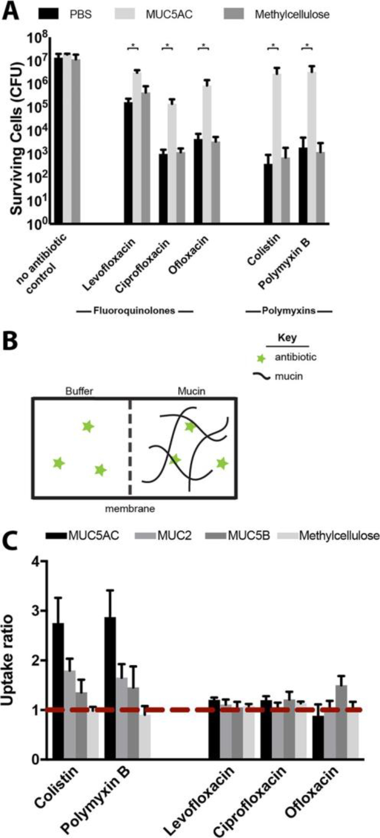
Evaluating the mechanism of mucus/mucin reduction of antibiotic efficacy. Although MUC5AC (0.5% w/v) protected bacterial cells from killing compared to buffer alone, (A) methylcellulose (0.5% w/v) did not, suggesting that neither mechanical cues nor macromolecular crowding are responsible for the protection observed. All error bars represent standard deviation of biological replicates (n ≥ 3). (*) indicates a significant increase in the number of surviving cells after antibiotic exposure in mucus compared to that in mucin-free buffer (PBS), as determined by the t test (P < 0.01) (B) Schematic illustration of experimental set up for equilibrium dialysis experiments to evaluate polymyxin and fluoroquinolone binding to mucin (C) Uptake ratio from equilibrium dialysis for each antibiotic with MUC5AC, MUC2, MUC5B and methylcellulose, in the conditions used to evaluate antibiotic efficacy. Ratios greater than 1 (red dotted line) indicate binding. Colistin and polymyxin B both bind to MUC5AC, MUC2, and MUC5B but not to methylcellulose. Uptake ratios of fluoroquinolone antibiotics to biopolymers are close to 1, consistent with weak or no binding. Error bars represent standard error of biological replicates (n ≥ 3) (see Methods for standard error calculation).