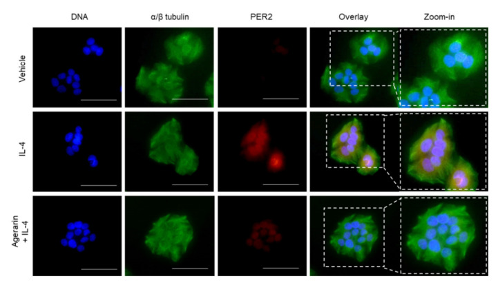
Figure 5.
Effect of agerarin on IL-4-induced localization of PER2. HaCaT cells cultured on coverslips were treated with vehicle (PBS), 20 ng/mL IL-4, or 20 ng/mL IL-4 plus 40 μM agerarin for 24 h, followed by fixing and permeabilization. Immunofluorescence staining was performed using anti-PER2 and Alexa Fluor 555-conjugated secondary antibodies (red staining). The α/β-tubulin was counterstained with anti-α/β-tubulin and Alexa Fluor 488-conjugated secondary antibodies (green staining). Nuclear DNA was stained with 1 μg/mL Hoechst 33258 (blue staining). Fluorescent cells were captured with an EVOSf1 fluorescence microscope. The last panel on the right is a zoomed-in view of the dotted box. Scale bars, 50 μm.

