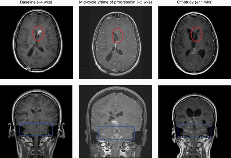
Figure 2.
Patient 3 serial gadolinium-enhanced T1 brain MRIs. Gadolinium-enhanced T1 MRI images acquired at Baseline, Time of Progression, and Off-Study for patient 3. Top series of images represents supratentorial axial slices. Bottom series of images represents coronal slices. Lighter shades within circle or square represent enhancing tumor. Transient regression of left periventricular lesion marked by circles in upper row. Progressive brainstem disease marked by rectangles in lower row. Blinded assessment performed by neuroradiologist (D.R.J.).

