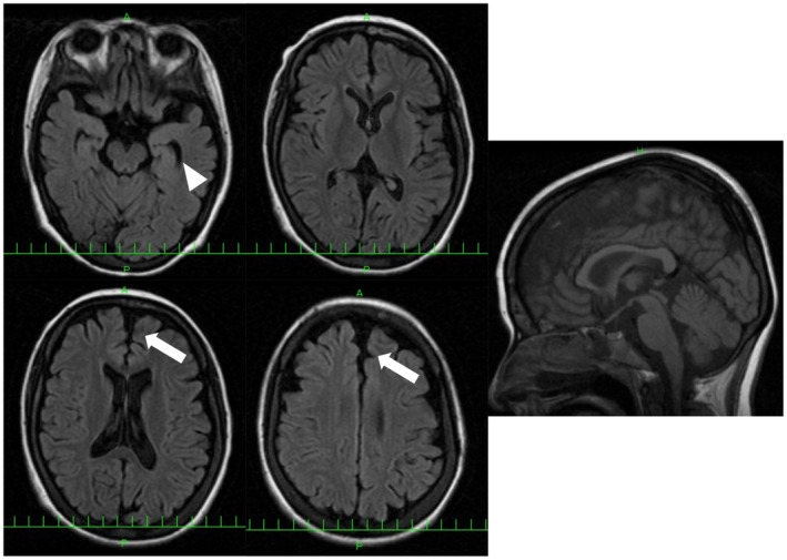
Figure 3.

Brain MRI of patient 7 with p.Tyr679Ser variant. FLAIR images show frontal (arrow) and temporal lobe atrophy (arrow head). White matter change and corpus callosum change are not observed. [Colour figure can be viewed at wileyonlinelibrary.com]
