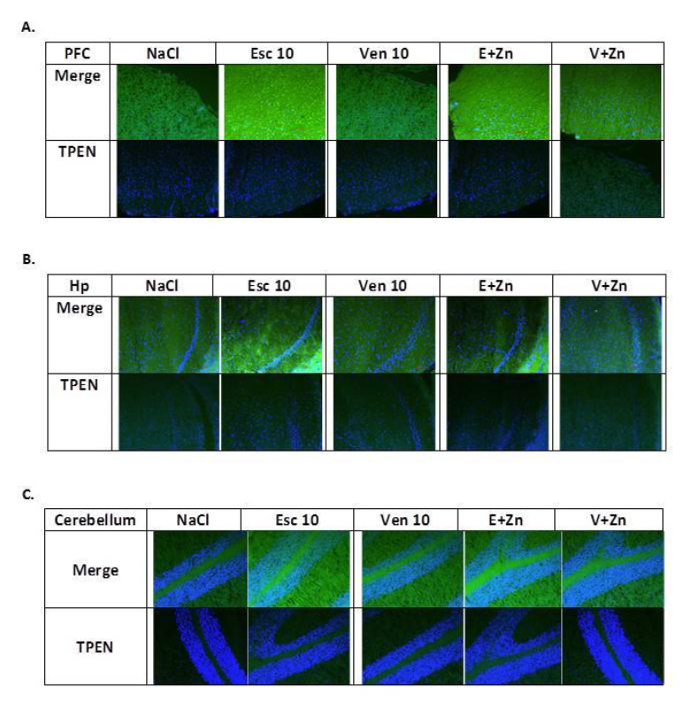
Figure 3.
The representative images of the effects of antidepressants (Esc, Ven) and Zn supplementation on intracellular zinc concentration in the prefrontal cortex (PFC, (A)) and hippocampus (Hp (B)), and cerebellum (C) of rats subjected to the OB + ZnD model using fluorescent Zinpyr-1 (ZP-1) staining. Sections were double-stained with DAPI (blue) to reveal cell nuclei and ZP-1 (green) to detect fluorescent intracellular Zn2+. Merged images include ZP-1 and DAPI (upper panel) or TPEN/ZP-1 and DAPI (lower panel) in the PFC (A), Hp (B), and cerebellum (C). ZP-1 fluorescence is mostly absent in TPEN-treated sections, indicating high specificity of ZP-1 for Zn2+. Scale bar = 50 μM.

