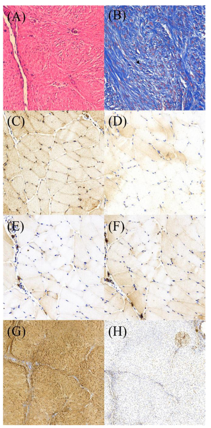
Figure 11.
Photos of histology and IHC stain of the specimens from repaired tendons. (A) H&E stain (400×). (B) tenocytes (arrow) by Masson’s trichrome stain (400×). IHC stain for growth factors (800×): (C) VEGF, (D) BMP-2. (E) TGF-B, and (F) vWF. (G) and (H) showing collagen I and collagen III under IHC stain (400×).

