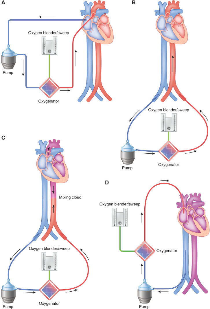
Figure 5.
ECMO may be configured either peripherally or centrally with different risks and benefits of either choice. (A) Diagram demonstrating central cannulation for VA ECMO. Right atrium/Inferior Vena Cava (IVC) is cannulated, and blood is flowing to the ECMO pump into the oxygenator and flowing back into the patient in an antegrade fashion directly into the ascending aorta. (B) Diagram demonstrating peripheral cannulation for VA ECMO. The femoral vein is cannulated, and blood is flowing from the patient to the ECMO pump into the oxygenator and flowing back into the patient in a retrograde fashion via the femoral artery. For (A) and (B), the oxygen blender and sweep as shown can be titrated accordingly on the basis of the partial pressure of oxygen in arterial blood (PaO2) and the partial pressure of carbon dioxide in arterial blood (PaCO2). This is the most invasive type of cannulation for ECMO. (C) Diagram demonstrating North-South syndrome or Harlequinn syndrome as seen in patients with peripheral VA ECMO. The mixing cloud occurs at the junction of retrograde oxygenation blood from the ECMO with that of the patient’s native LV cardiac output. Blood proximal to the mixing cloud is often deoxygenated, hence the importance of a right upper extremity arterial line in peripheral VA ECMO. (D) Diagram demonstrating VV ECMO with a right internal jugular vein and right femoral vein cannulation. Blood is flowing from the right femoral vein to the ECMO pump into the oxygenator and returning back into the patient in the right internal jugular vein.

